Large grant for research in computer methods for fighting diseases
Centre for Stochastic Geometry and Advanced Bioimaging (CSGB), which this department is involved in. At DIKU, the grant will be used to complete 3 specific projects within the next 5 years: Mapping the pathways in the brain, modeling how brains affected by dementia change over time, and creating visualizations of neuronic vesicles.

The Villum Foundation has recently decided to support the Danish Centre for Stochastic Geometry and Advanced Bioimaging (CSGB), with 30M DKK during the next 5 years. The centre was established in 2010 with a previous Villum Foundation grant of 25M which initiated a collaboration between computer scientists, mathematicians, statisticians, biophysicists and molecular biologists from research groups at Aarhus University, Aalborg University and the Department of Computer Science at University of Copenhagen (DIKU). From the outset the ambition of CSGB has been to develop a new generation of methods for analyzing advanced biological image data – and this point of departure has not changed even though the projects planned for the center’s second period are new.
The expertise of computer scientists is essential
At DIKU, scientists have great expertise in extracting important information from medical images, which can be useful in the healthcare industry. This accumulated expertise will be essential for several interesting research projects: 1) mapping the pathways of the brain, 2) determining how the brain changes appearance as dementia progresses, and 3) study the movements of neurotransmitters.
The common denominator for all the projects at DIKU are that they intersect computer science, mathematics, and statistics and that they use image data as input. The strength of the computer scientists is that they can use the computer as a tool for developing advanced mathematical and statistical models, which can help make sense of the big data sets of medical images.
DIKU scientists are mapping fluids path through the brain
7 tenured researchers and 5-6 PhDs and postdocs from DIKU will be involved in the new stage of the centre’s activities.
During the next 5 years, Aasa Feragen and François Lauze are plunging into the neurobiological discipline, tractology, to study how the regions of the brain are connected. Using diffusion MRI scans of the brain, the scientists are able to model the direction of fluid movements in the brain and thus infer how the neurons are connected. Apart from adding to our knowledge of how the brain works, tractology have concrete applications in connection with e.g. brain surgery, providing the doctors with important information about which centers of the brain are connected and thus where they must be especially careful.

Diffusion MRI scans of brain tissue. The colored areas of white substance have automatically been detected using computational analysis of the picture.
Early diagnose of Alzheimer’s disease
The work of Stefan Sommer is concerned with Alzheimer’s disease – a debilitating illness that the health care industry has great interest in trying to prevent. Alzheimer’s disease progresses over a long period of time and it is therefore a serious challenge to discover it in time. Furthermore, many years (10-15 years) pass before it is possible to measure the effect of preventive treatment.
Stefan’s project involves creating statistical models which can be used for discovering indicators af Alzheimer’s earlier than it is possible today. Using data from MR scans it is possible to "create models of a brain diagnosed with Alzheimer’s in all of stages of the disease as well as models of a healthy brain for comparison. Thus, the computer will be able to recognize early indicators of the disease, which are difficult for the human eye to perceive.
How does stress effect our brain functions?
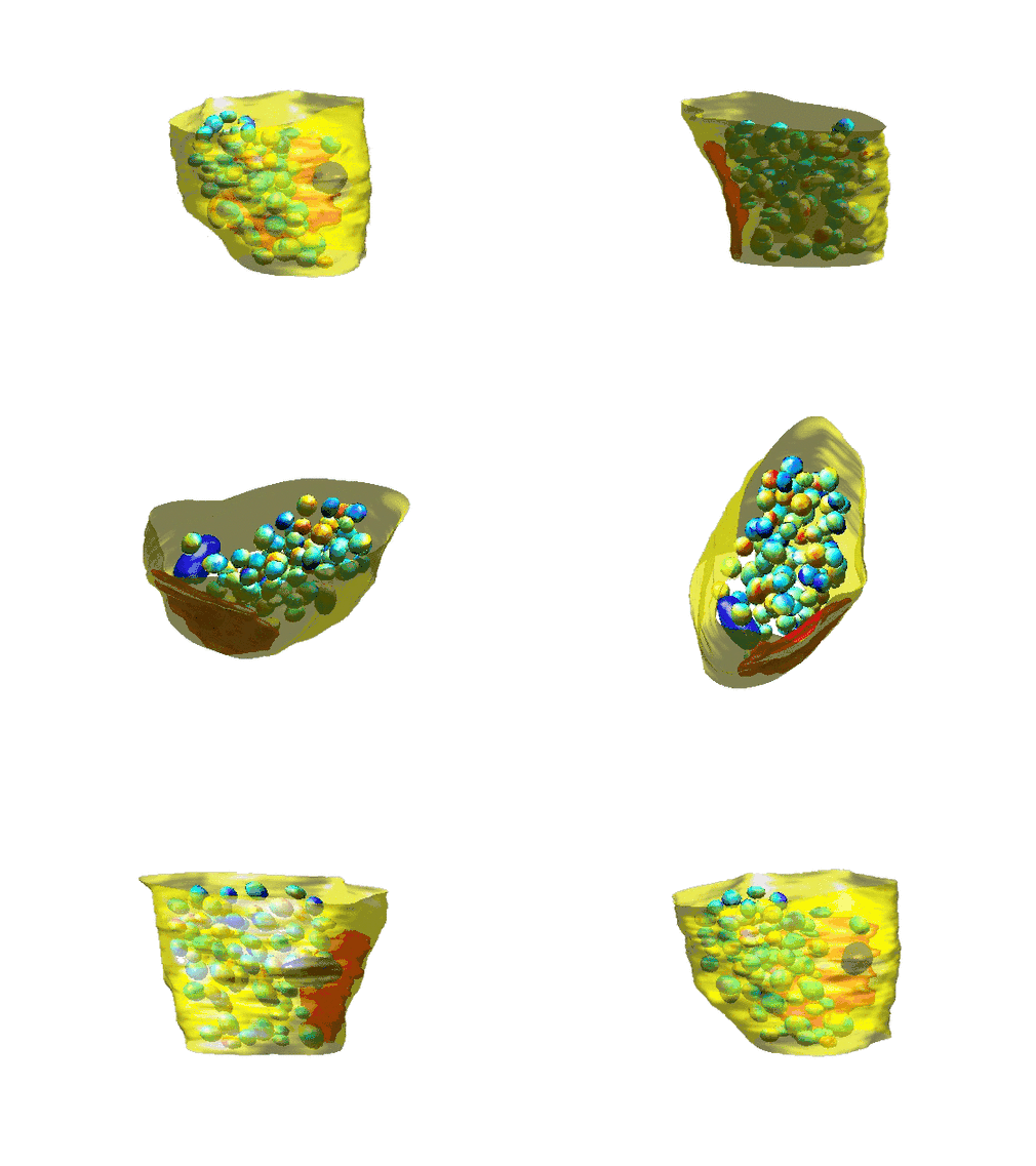
Jon Sporring is involved with CSGB with a project about the effect of stress on the brain. The computer scientists are able to detect how the neurons of the brain are connected by studying the neurotransmitters through electron microscopy – more specifically the very detailed 'Focused Ion Beam Scanning Electron Microscopy (FIB-SEM)', which makes it possible to cut the brain tissue into 5 nanometer thin slices and take pictures of them. The computer scientists make use of the information in the pictures about the position and form of the vesicles to create a 3D visualization. By comparing the visualization of vesicles created from brain tissue of lab rat that have been exposed to stress and visualizations generated from stress free rats, it is possible to see how stress affects the nerves.

Visualization of the brain cells' (the synapses’) vesicles and their content of neurotransmitters.
