
BioImaging
Bioimaging collectively refers to tools for creating and studying structural or functional images of living objects and systems.
At DIKU we use the term bioimaging for the study of statistics and geometry of 3-dimensional structures observed through microscopes. Examples are
- Tomographical reconstruction of protein structures from cryo-electron microscope images
- Estimating the density of synaptic vesicles in the brain of rats
- Modelling of nano-domains on plant surfaces.
Our projects are realized in close collaboration with experts from the fields of molecular biology, microscopy, statistics and mathematics, and our prime collaborators are the Center of Stochastic Geometry and Advanced Bioimaging (CSGB) as well as the national instrument center, Center for Advanced Bioimaging (CAB).
Vesicle modelling
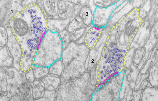
The image shows a serial section transmission electron microscope (ssTEM) image, where 3 synapses have been annotated. The yellow and cyan denote regions of the pre- and post-synaptic compartments, the blue circles are vesicles, and purple is the active zone, where vesicle release their neurotransmitters.
In this example, we are interested in, whether the 3-dimensional distribution of vesicles as a function of the shortest distance to the active zone differs between stressed and non-stressed animal models.
Tomography of cryo-electron microscopy images
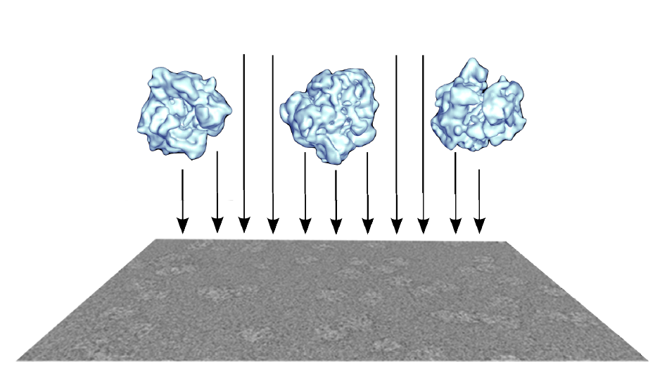
We study the structure of proteins by Single-particle reconstruction (SPR), where thousands of similar copies of proteins in random orientations are imaged by transmission electron microscopy.
The central problem is to perform the 3-dimensional reconstruction of the protein mass density from a set of 2-dimensional images with unknown orientation and protein configuration. The image illustrates how different molecules are projected onto an image.
People
More researchers coming soon
| Name | Title | Phone | |
|---|---|---|---|
| Christian Igel | Professor | +4535335674 | |
| Jon Sporring | Professor | +4524252334 |
Contact
 Jon Sporring
Professor
sporring@di.ku.dk