Medical Imaging
The Medical Image Analysis lab is concerned with the analysis of images for medical purposes. The lab is focused on the quantification of pathological changes through medical imaging biomarkers.
The major applications are
- Neuro Imaging
- Breast cancer screening
- Pulmonary images
The lab is focused on the quantification of pathological changes through medical imaging biomarkers.
Neuro Imaging
Within neuroimaging some of our projects include longitudinal changes in Alzheimer’s patients and the quantification of the disease progression in collaboration with Glostrop Hospital. We collaborate with Institute for Neuro Pharmacology at the faculty of Health on projects concerning plasticity of the brain with focus on blind subjects and with the Coma Science Group in Liege on the pattern of conscious awareness in coma patients.
The medical imaging lab is working with the processing PET images with focus on artifact removal Attenuation correction and image-based normalization for quantification. This work is in collaboration the Copenhagen University Hospital, Dep. of Clinical Physiology, Nuclear Medicine and PET
Breast Cancer Screening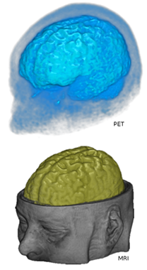
Using the state of the art image analysis and machine learning techniques the image group is involved in a HTF project on breast cancer screening.
The goal is to develop an imaging biomarker, which can aid in the early detection of breast cancer.
Pulmonary Imaging
COPD is a major cause of death. This project focuses on developing imaging biomarkers for the progression of the disease in collaboration with Gentofte hospital.
Projects
Breast cancer, osteoporosis and hardening of the arteries are all widespread diseases where an early diagnosis can be a matter of life and dead.
The Learning Imaging Biomarkers research project aims to create more effective diagnostic tools by designing efficient biomarkers. The researchers work with the extraction of shapes and appearances from mammograms and x-rays and with the development of geometrical models that allow similarity measures to be computed from them. The similarity measures are used to train learning machines to recognise biomarkers being much more reliable than traditional markers. The result is a more accurate patient diagnosis, prognosis and prediction of disease progression.
The researchers are collaborating with clinics performing traditional screenings around data and a biological understanding developed from experience with x-rays.
- Cardiovascular disease (CVD)
Atherosclerosis is usually the underlying pathology of cardiovascular diseases (CVD) and CVD causes 58 % and 48 % of all death in United States and Europe, respectively. - Breast Cancer Risk Assessment
The project is a collaboration between DIKU and Nordic Bioscience Imaging funded by a combination of DIKU, the LImB project, grundforskningsfonden, the research councils, and Nordic Bioscience Imaging. - Osteoarthritis (OA)
The project is a collaboration between DIKU and Nordic Bioscience Imaging funded by a combination of DIKU, the LImB project, Grundforskningsfonden, the research councils, and Nordic Bioscience Imaging. - COPD
The project is a joint initative of the Image Group at University of Copenhagen and Gentofte University Hospital in Copenhagen. Chronic obstructive pulmonary disease (COPD) is a major cause of morbidity and mortality worldwide. - Shape classification and quantification in medical image analysis
The aim of the project is to create a mathematical framework for analysing treelike shapes in images, and in particular medical images - for instance in connection with vascular calcifications, smoker's lungs or cancer vascularization.
People
| Name | Title | Phone | |
|---|---|---|---|
| Christian Igel | Professor | +4535335674 | |
| Francois Lauze | Associate Professor | +4535335671 | |
| Jon Sporring | Professor | +4524252334 | |
| Sune Darkner | Professor | +4535335702 |